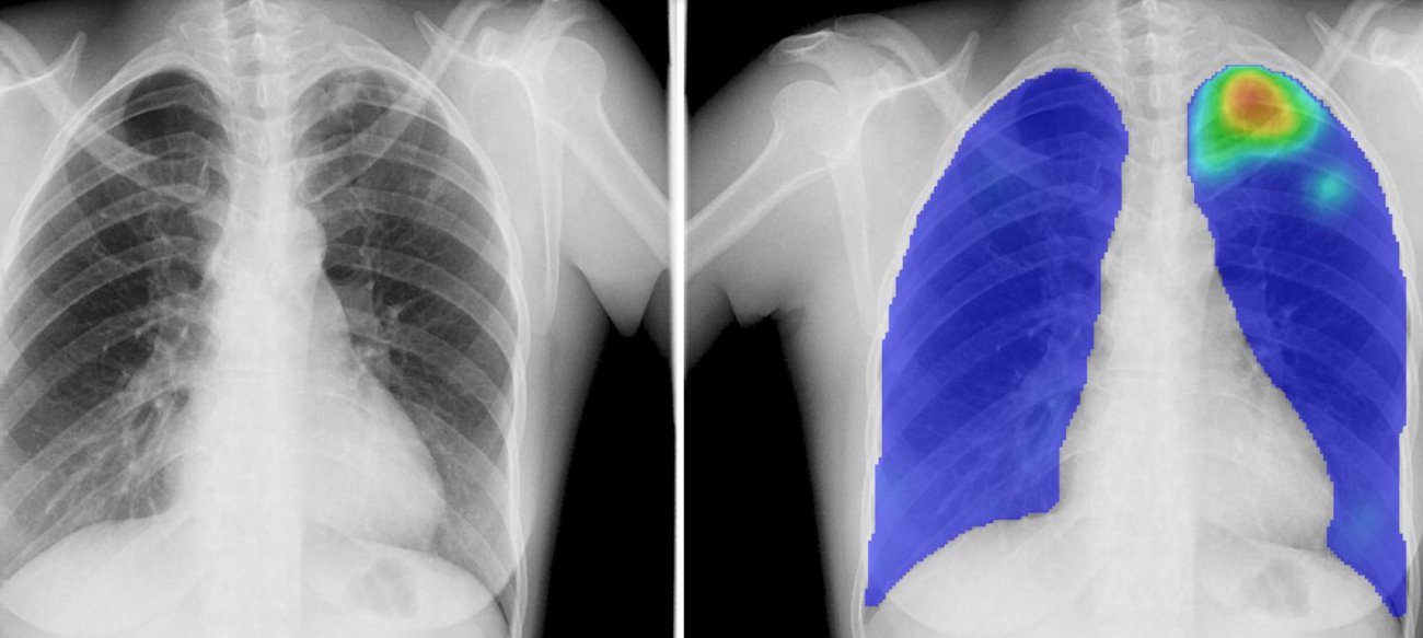
A CT scan takes a series of photos of cross-sectional areas of a body and combines them into a 3D image. A radiologist must look at each section to see if there are any abnormalities. This is a time-consuming task for highly trained healthcare staff, of which there is a shortage. Bram van Ginneken, Professor of Medical Image Analysis at Radbouumc, is working on automating this assessment. In recent years, we have discovered that large neural networks made up of many layers - which is why we call them deep learning - can be used to achieve excellent results when interpreting a medical image.
Continuous training
We have developed and trained algorithms with tens of thousands of examples of CT scans. On these, doctors had indicated which abnormalities could indicate lung cancer,' says Van Ginneken. Then the system analysed the new CT scans. Doctors checked those assessments again and improved or supplemented them if necessary. ‘In the end, we created a system that can assess scans at the population level just as well as doctors can.’ The use of these systems helps doctors to detect conditions at an earlier stage, which in turn increases the chances of successful treatment. However, the algorithm does not yet succeed in recognising rare cases that it has never seen before. ‘So it is crucial that we continue to train such an artificial intelligence-based system with new data.'
Cyclical process
Van Ginneken has been working for years on the automatic detection of abnormalities on CT scans. His Vici project, which started in 2013, led to software that supports radiologists in analysing scans and is now used worldwide. A long haul is needed here: medical innovations often take up to fifteen years before a product is actually on the market. Good cooperation with end users such as radiologists is of great importance. ‘It's a cyclical process,' explains Van Ginneken. ‘We develop software, the radiologists test it and immediately tell us what is not good about it. Then we improve it again and deploy it in a number of hospitals. You need those iterations to make a really good product.' In his Perspectief programme DLMedIA: Deep Learning for Medical Image Analysis, Van Ginneken also works together with various parties, such as Philips, to arrive at the next generation of image processing systems. 'Because we are here at a hospital and have direct access to the data and medical analysis, we can develop systems that can actually be used in the real world.'
Developing countries
Besides the increasingly advanced deep learning systems, Van Ginneken also works on low-cost healthcare for developing countries. ‘There, people are very happy if there is help to automatically assess X-rays for tuberculosis screening, for example'. In those countries there are simply no radiologists to analyse the photos. The software offers a solution there: no people need to be trained.
Future
There are now almost 200 products on the market that can support radiologists with the help of AI. But these are not yet being used very much. When that happens, we can really hand over certain tasks to the computer. Then the same number of people can continue to assess the increasing number of medical scans in less time,' says Van Ginneken.